Saturday, December 22, 2012

Arthroscopic Repair of Concomitant Type II SLAP Lesions in Large to Massive Rotator Cuff Tears

Arthroscopic Repair of Concomitant Type II SLAP Lesions in Large to Massive Rotator Cuff Tears
This is a very good study that supports the idea that the LH biceps tendon can be a significant pain generatory.  My personal experience is that a great deal more stiffness is seen in SLAP/RCR group and more pain....so my personal experience mirrors this groups findings.

Dr.  William D. Murrell M.D., M.Sc.
Consultant Orthopaedic Sports Medicine
Dubai Bone and Joint Center
Dubai, United Arab Emirates.

Wednesday, December 19, 2012

Adolescent athletes show asymmetric function in knee after ACL reconstruction | Orthopedics

Adolescent athletes show asymmetric function in knee after ACL reconstruction | Orthopedics

Even in a well reconstructed knee, the knee will probably have functional differences as compared to the unoperated side.

Dr. William Murrell
Consultant Orthopaedic Sports Medicine
Dubai Bone and Joint Center
Dubai UAE

Thursday, November 15, 2012

No significant difference found between drilling, grafting during ACL reconstruction | Orthopedics

No significant difference found between drilling, grafting during ACL reconstruction | Orthopedics

This research mirrors outcomes seen elsewhere.  However, the magic time period after this surgery utilizing drilling will probably see decline in the outcomes after 3-5 year time frame...will be interesting to see the outcomes at the 5 year mark.  I think that there will be a significant decrease in survivability at this time period.

Two-stage Revision Anterior Cruciate Ligament Reconstruction: Indications, Review, and Technique Demonstration | Orthopedics

Two-stage Revision Anterior Cruciate Ligament Reconstruction: Indications, Review, and Technique Demonstration | Orthopedics

This is an interesting approach, at DBAJ we like to also complete a one stage if possible, but two stage when bone stock does not allow placement of graft in optimum position. 

Wednesday, November 7, 2012

Cutting Edge Orthopaedic Biological Treatments for the 21st Century.....My latest article.
http://viewer.zmags.com/publication/853b88fd#/853b88fd/70

Sunday, October 21, 2012

Inter- and Intraobserver Reliability of the Radiographic Diagnosis and Treatment of Acromioclavicular Joint Separations | Orthopedics

Inter- and Intraobserver Reliability of the Radiographic Diagnosis and Treatment of Acromioclavicular Joint Separations | Orthopedics

Poor agreement with type of injury, whether to operatate, and how to fix.....We are still confused.

Two-year follow-up shows efficacy of endoscopic repair for gluteus medius tears | Orthopedics Today

Two-year follow-up shows efficacy of endoscopic repair for gluteus medius tears | Orthopedics Today

This may be a promising technique of glut medius tear repair.

Three lessons from sports applied to business | LinkedIn

Three lessons from sports applied to business | LinkedIn

Right from my playing days as a junior in the Indian Hockey team, I used to keep a small green diary with me (this was pre-mobile, pre-tablet era) at all times. I would write my experiences, learnings and motivational quotes which would keep me going during tough times. Today in the midst of the corporate world as the CEO of Olympic Gold Quest, stories in this diary still make so much sense and I would like to share these anecdotes with you: the Top 3 in my Green Diary. 
1. Lack of funds does not hamper work, lack of dedicated workers does - Jamnalal Bajaj
Vijay Kumar is one shooter we greatly respect at OGQ. We have been supporting him since 2010, and not once did I hear him grumbling about lack of funds for training or shortage of ammunition. He never complained about a cramped training schedule or lack of quality support staff for the Indian team. He did what he knew best: train like a disciplined army man. This pistol shooter quietly went about accomplishing his task of winning the silver medal at the London Olympics. I know so many athletes who always crib for the best equipments, kit, etc and yet when it comes to delivering at the grand stage, they fail. 
Similarly in corporate life, I advise you to make the best of the resources you have and focus on the task. Results will follow.      
      
2. Whether you think that you can, or that you can’t, you are usually right - Henry Ford
Mary Kom is the most inspirational lady I have ever met in my life. She came from a village in Imphal where people -- including her family -- were against girls taking up boxing. She continued to fight and went on to win multiple world championships. Mary took a sabbatical from sports for 2 years in 2006. She got married, delivered twins and came back to win 2 more world championships. OGQ signed her in 2009 and she went on to win her fifth world championship in 2010. She was 28 by then and many people thought she was past her prime. Mary had other thoughts. OGQ made a detailed plan with Mary Kom keeping in mind that for the London Olympics, Mary would have to shift from 46kg to 51kg. We identified a world class foreign coach and physio for her and she gave her 200% in the training camps. If anyone had the will, determination and grit to win an Olympic medal for India, it was Mary Kom. I salute the courage and perseverance of Mary Kom.
Many times you will feel like giving up on your job or scared to take a risk. Mary is a shining example of converting criticism into inspiration and always believing in yourself. 

3.       You must be the change you want to see - Mahatma Gandhiji
This is the motto we have adopted at OGQ. The easiest thing to do about the Indian sport scene is to complain: "A population of 1.2 billion and just handful of Olympic medals". We realized early that there are lot of hurdles and problems in our country. The best we could do was to focus on the good things and ensure our athletes got the support they deserved. We worked on plugging the gaps in the preparation of our athletes, rather than complaining and grumbling on the problems ailing the sports structure. We believed that sports in India will grow once young people have good role models to follow. Six Indian athletes won medals at the London Olympics (a record haul for India) and 4 are supported by OGQ. This is a watershed moment in the history of Indian sports. Kids across the country are inspired to take up sports. Parents believe their kids can make a career in Olympic sports. Government is pumping more funds and federations are getting more professional. There is still a lot of room for improvement, but we believe we have played a small role in this big change.
Companies, mostly big ones, sometimes hesitate to make changes. For young professionals it can become suffocating to work in such an environment. But, instead of complaining about the system, go and be the change. Show people the pros of a change and systems will change for good.

Tuesday, October 9, 2012

ACL lesions, lateral meniscal tears correlated with increased tibial plateau fractures | Orthopedics

ACL lesions, lateral meniscal tears correlated with increased tibial plateau fractures | Orthopedics

ACL lesions, lateral meniscal tears correlated with increased tibial plateau fractures

  • October 4, 2012
Increased tibial plateau fracture depression shows a significant impact on ACL lesions and lateral meniscal tears, according to this study.
“Articular depression is a potential predictor of specific meniscal and ligamentous injuries in acute tibial plateau fracture,” Alexander S. Spiro, MD, and colleagues stated in the abstract. “Magnetic resonance imaging is generally recommended with respect to associated soft tissue injuries, especially in cases with distinct tibial plateau fracture depression on multi-detector computer tomography scans.”
Fifty-four consecutive patients with acute tibial plateau fractures were admitted to a university clinic’s emergency department. The amount of articular depression was measured with a multi-detector computer tomography, according to the abstract.
Spiro and colleagues then compared the scans with the patients’ MRI images for meniscal tears, crucial and collateral ligament injuries and patellar retinaculum lesions. After logistical regression, they found increased tibial plateau fractures correlated with incidence of meniscal lateral tears and ACL lesions, while analysis of covariance showed that the amount of articular depression was significantly associated with the absolute number of soft tissues injuries, according to the abstract.

Friday, September 28, 2012

The Effect of Smoking on Ligament and Cartilage Surgery in the Knee

The Effect of Smoking on Ligament and Cartilage Surgery in the Knee

The Effect of Smoking on Ligament and Cartilage Surgery in the Knee

A Systematic Review

  1. David C. Flanigan, MD*,
+ Author Affiliations
  1. *The Ohio State University Sports Medicine Center and Cartilage Restoration Program, Columbus, Ohio
  2. Department of Orthopedic Surgery, Washington University School of Medicine, St Louis, Missouri
  3. Penn Center for Advanced Cartilage Repair and Osteochondritis Dissecans Treatment Perelman School of Medicine at the University of Pennsylvania, Philadelphia, Pennsylvania
  4. §Department of Orthopaedic Surgery and Sports Medicine, University of Kentucky, Lexington, Kentucky
  5. Investigation performed at The Ohio State University Sports Medicine Center, Columbus, Ohio
  1. David C. Flanigan, MD, The Ohio State University Sports Medicine Center, 2050 Kenny Rd, Suite 3100, Columbus, OH 43221-3502 (e-mail: david.flanigan@osumc.edu).

Abstract

Background: The adverse effects of smoking on various health conditions such as cancer, diabetes, and cardiovascular disease have been well documented. Many orthopaedic conditions, such as fracture healing, wound repair, and bone mineral density, have been reported to be adversely affected by smoking. However, no known systematic reviews have investigated the effects of smoking on ligament and cartilage knee surgery.
Purpose: We hypothesized that smoking would have a negative influence from both a basic science and clinical outcome perspective on these types of knee surgeries.
Study Design: Systematic review.
Methods: A systematic review of multiple medical databases was performed evaluating clinical and basic science studies to determine the effects of smoking on ligament and cartilage knee surgery.
Results: Fourteen studies were found for inclusion and analysis. Eight of these studies addressed the relationship between smoking and knee ligaments, and 6 investigated the relationship between smoking and articular cartilage. With the exception of 1, all of the basic science and clinical studies exploring the relationship between smoking and knee ligaments found a negative association of smoking, either molecularly, biomechanically, or clinically. One basic science and 3 clinical studies found a negative influence of smoking on articular cartilage of the knee. No studies were found that investigated the relationship of smoking and menisci.
Conclusion: The current literature reveals a negative influence of smoking on the results of knee ligament surgery, both from a basic science and clinical perspective, implying that smoking cessation would benefit patients undergoing these procedures. The association between smoking and knee articular cartilage was less clear, although the literature still suggests an overall negative influence and highlights the need for further investigation.

Keywords:

Monday, September 24, 2012

New hydrogel may help cartilage regeneration research | Orthopedics

New hydrogel may help cartilage regeneration research | Orthopedics

New hydrogel may help cartilage regeneration research

  • September 24, 2012
A new synthesized hydrogel may help replace lost cartilage in joint replacement patients with osteoarthritis, according to Harvard researchers.
“For a gel to work in those settings, it has to be able to stretch and expand under compression and tension without breaking,” Jeong-Yun Sun, a postdoctoral fellow at the Harvard School of Engineering and Applied Sciences, stated in a press release.
Using a combination of polyacrylamide and alginate, the synthesized gel can stretch up to 21 times its size and is stronger than gels formed by either material alone, researchers said. Compared to other hydrogels often used as scaffolds for cartilage regeneration, this new “super gel” maintains its mechanical stability and elasticity over multiple stretches.
Sun and colleagues found that, even after the gel cracked, it was able to stretch to 17 times its own size. The researchers said they hope the gel will be an alternative to more invasive-based approaches, such as autologous chondrocyte implantation or joint replacement, in the treatment of patients with osteoarthritis.

A solution to reducing inflammation

A solution to reducing inflammation

Contact: Morwenna Grills
Morwenna.Grills@manchester.ac.uk
44-016-127-52111
University of Manchester

A solution to reducing inflammation

Research carried out at The University of Manchester has found further evidence that a simple solution, which is already used in IV drips, is an effective treatment for reducing inflammation.
The researchers also identified that hypertonic solution, which is a solution with an elevated concentration of salt, can ease inflammation purely through bathing in it – proving the Victorians were right to visit spa towns to "take the waters" for ailments like rheumatoid arthritis.
The research team, led by Dr Pablo Pelegrin, was investigating how cell swelling can control inflammation; the immune system's first response to injury or infection.
They discovered that white blood cells swell in a similar way to how tissue swells around a wound. The team then went on to look at what causes the swelling.
The researchers injected solutions with low ions into mice. They found that these solutions acted as a danger signal, causing cells to swell. The swelling then activates a group of proteins called NLRP3 which then release inflammatory mediators. These activate neighbouring cells to increase inflammation.
However, when a hypertonic solution was administered to the mouse it drew the water out of the cells shrinking them back to their original size. This in turn deactivated the signal for inflammation.
Dr Pelegrin's research provides further evidence for the use of hypertonic fluid therapy for the reduction of inflammation in the brain, a treatment that can reduce the amount of damage caused by illnesses such as stroke and epilepsy. His team has been able to show for the first time why the solution works at a molecular level.
Dr Pelegrin says: "Hypertonic solutions have been used in the treatment of stroke for many years. Clinicians have found that their use not only reduces brain swelling, but also alleviates brain inflammation. However, because there wasn't a molecular target for hypertonic solutions there has been a lot of debate about the clinical effect. Here we have indentified a target for hypertonic solutions by blocking the NLRP3 inflammasome which triggers inflammatory mediators at a molecular level".
The team also looked at the benefits of hypertonic solutions when used outside of the body. They soaked bandages in the solution before using them on the legs of mice. They also tested bathing the inflamed area in a hypertonic solution and in both cases the inflammation was reduced.
It appears the hypertonic solution produces an osmotic gradient through the skin, which explains why hot springs, which have a hypertonic make up, can ease the pain of conditions such as rheumatoid arthritis.
Vincent Compan worked with Dr Pelegrin on this research in the Faculty of Life Sciences. He says: "This research opens up exciting opportunities for the use of hypertonic solution as a treatment for inflammatory illnesses such as arthritis. What we've identified has the potential to be used to help so many patients."
Another aspect of the team's research identified that the signalling process to activate inflammation is one of the oldest evolutionary processes. The researchers found that the same mechanism of cell swelling causes NLRP3 inflammasome activation in fish as well as mammals. This means it is one of oldest responses in the body leading to inflammation.
The research has recently been published in the journal Immunity.

Anti-inflammatory cytoki... [Knee Surg Sports Traumatol Arthrosc. 2012] - PubMed - NCBI

Anti-inflammatory cytoki... [Knee Surg Sports Traumatol Arthrosc. 2012] - PubMed - NCBI

Knee Surg Sports Traumatol Arthrosc. 2012 Sep 15. [Epub ahead of print]

Anti-inflammatory cytokine profile in early human tendon repair.

Source

Integrative Orthopedic Laboratory, Department of Molecular Medicine and Surgery, Karolinska Institutet, Stockholm, Sweden, paul.ackermann@karolinska.se.

Abstract

PURPOSE:

The aim of this study was to assess inflammation and the presence and relative levels of cytokines, which may be involved in regulating early human Achilles tendon healing.

METHODS:

Nine patients with acute Achilles tendon rupture were included, operated on and post-operatively immobilized. Two weeks post-operatively, microdialysis of the peritendinous interstitial compartment was performed in the healing and intact contralateral Achilles tendons. Quantification of tumour necrosis factor (TNF)-α, interferon (IFN)-γ, interleukin (IL)-1β, IL-6, IL-8, IL-10, IL-12p70 and IL-17A was accomplished using a cytometric bead array. Prostaglandin (PG) E(2) levels were measured by enzyme immunoassay.

RESULTS:

None of the patients displayed detectable PGE(2) levels. Pro-inflammatory cytokines were below detection levels (IFNγ, IL-12, and IL-17) or did not differ between injured and control tendons (IL-1β and TNF). Notably, IL-6, IL-8 and IL-10 concentrations in the healing Achilles tendon were significantly elevated: 13-fold (p = 0.009), 28-fold (p = 0.02), and 3.7-fold (p = 0.03), respectively.

CONCLUSION:

At 2 weeks post-human Achilles tendon rupture, healing is characterized by a resolving inflammatory phase and up-regulation of IL-6, IL-8 and IL-10. The absence of inflammation suggests that at this time point, these cytokines may be associated with anti-inflammatory and regenerative effects on the tendon healing process.

Wednesday, September 12, 2012

Family history, gender affect survival of ACL graft at 15-year follow-up

Family history, gender affect survival of ACL graft at 15-year follow-up

Bourke H. Am J Sports Med. 2012. doi:10.1177/0363546512454414.

  • September 12, 2012
Patients who underwent primary ACL surgery 15 years ago report an 86% or higher survival rate for their graft at latest follow-up, according to this study.
Surgeons performed a single-incision endoscopic technique using either an autologous bone-patellar tendon-bone graft (BPTB) or hamstring tendon (HT) graft in 755 patients. At minimum 15 years follow-up, 689 patients completed a subjective questionnaire. The mean International Knee Documentation Committee subjective score was 85 at 15 years and 73% of patients reported return-to-sport preinjury levels, with 51% still performing strenuous or very strenuous activities, according to the abstract.
“Anterior cruciate ligament reconstruction using this technique is a reliable and reproducible procedure when using either the BPTB or HT autograft and allowed 73% of patients a return to their preinjury sports with an ACL graft rupture rate of less than 1% per year,” Henry E. Bourke, FRCS (Tr&Orth) stated in the study.
After the 2-, 5-, 10- and 15-year follow-up, expected survival of the ACL graft was 95%, 93%, 91% and 89%, respectively, according to the abstract. Expected survival of the contralateral ACL graft was 97%, 93%, 90% and 87% during the same respective follow-up periods. Although graft choice did not affect the risk of rupture, contralateral ACL grafts were twice as likely to rupture in the BPTB group as in the HT group.
Researchers also noted that risk factors for rupture included male gender, which lowered the chances of survival, and a family history of ACL rupture, which doubled the chances of rupture in both the ACL and contralateral ACL groups.

Clinician calls for further study on platelet-rich plasma formulations and impact

Clinician calls for further study on platelet-rich plasma formulations and impact

  • Orthopedics Today, September 2012
MONTREAL — While there is evidence of successful treatment using platelet-rich plasma for various pathologies, the lack of unfailing positive clinical outcomes points to variability among patients and materials, according to a lecturer at the International Cartilage Repair Society World Congress 2012.
In his lecture, Scott A. Rodeo, MD, described his and others’ clinical experiences with platelet-rich plasma (PRP). His message — more research is needed about these materials and understanding the variability inherent in PRP formulations, patients and their pathologies is key.
“One of the fundamental issues, as a clinician if you are thinking of using PRP, is the tremendous variability,” Rodeo, of the Hospital for Special Surgery in New York, said. “All PRPs are not the same. There is variability between the various commercial systems that are available. There is also variability, of course, within the individual patient and the underlying biology.”
Scott Rodeo 
Scott A. Rodeo
He noted that cytokines influence several biological aspects of connective tissue.
“Clearly, connective tissue healing requires a complex timing and sequence of cytokine expression, so the rationale and attraction of PRP is the ability to deliver numerous cytokines in potentially physiologically relevant proportions,” Rodeo said. “It is complex, and it is a bit of a leap of faith. In fact, despite vast basic science and laboratory data [that] demonstrate a positive effect of PRP on these tissues, this has not translated into a consistently positive clinical effect.”

Variability

A reason for this, Rodeo suggested, could be that PRP formulations differ among manufacturers and patients. Even within each individual, formulations can vary throughout the day.
With these variations, it becomes impossible to determine whether a formulation that works in one study will have the same effect in another patient for a similar application or what may be the content of a given formulation.
“What are we putting in the patient? With a drug, it is easy. We know it is a precise composition,” Rodeo said. “That contrasts with the PRP materials we are using in patients.”

Arthritis treatment

Although a number of studies have demonstrated a clinical improvement in symptoms after PRP injections for the treatment of arthritis, Rodeo noted that most of the data available refer only to the knee.
“There is little data for other joints,” he said. “Most studies do report better results in younger patients with lesser degrees of degeneration. The clinical effect typically wears off in 6 months to 12 months. There is little data demonstrating a positive structural effect — that is, actual cartilage regeneration — so it may be symptom-modifying but it is probably not structure-modifying.”
Rodeo noted that more research is needed about PRP.
“We certainly need more information,” he said. “Keep in mind, arthritis is a heterogeneous condition. The effect of a specific PRP formulation may differ significantly based on the underlying biologic or inflammatory milieu.”
He added, “Clinical results, clearly, are mixed. PRP seems to have the potential to be symptom-modifying, but clearly [it] does not appear to be structure-modifying.”
With regard to the variability of PRP, Rodeo noted clinicians must strive to understand what they are delivering to the patient. This means analyses of PRP samples, so these formulations can be correlated with clinical results.
“We know cytokines can increase production of matrix proteins, but a critical deficiency with all this work is that tissue microstructure is not reformed,” Rodeo said. “We do not regenerate normal cartilage, tendon or meniscus. [It] appears growth factors, such as PRP, still do not provide the proper cellular and molecular signals to truly drive regenerative healing. I would submit that both cells and the signals are needed to reconstitute both tissue composition and structure.” – by Robert Press
Reference:
  • Rodeo SA. Clinical experience with platelet-rich plasma. Paper 15.1.3. Presented at the International Cartilage Repair Society World Congress 2012. May 12-15. Montreal.

Saturday, September 8, 2012

Highly stretchable and tough hydrogels : Nature : Nature Publishing Group

Highly stretchable and tough hydrogels : Nature : Nature Publishing Group

Highly stretchable and tough hydrogels

Nature
489,
133–136
(06 September 2012)
doi:10.1038/nature11409
Received
Accepted
Published online
Hydrogels are used as scaffolds for tissue engineering1, vehicles for drug delivery2, actuators for optics and fluidics3, and model extracellular matrices for biological studies4. The scope of hydrogel applications, however, is often severely limited by their mechanical behaviour5. Most hydrogels do not exhibit high stretchability; for example, an alginate hydrogel ruptures when stretched to about 1.2 times its original length. Some synthetic elastic hydrogels6, 7 have achieved stretches in the range 10–20, but these values are markedly reduced in samples containing notches. Most hydrogels are brittle, with fracture energies of about 10Jm−2 (ref. 8), as compared with ~1,000Jm−2 for cartilage9 and ~10,000Jm−2 for natural rubbers10. Intense efforts are devoted to synthesizing hydrogels with improved mechanical properties11, 12, 13, 14, 15, 16, 17, 18; certain synthetic gels have reached fracture energies of 100–1,000Jm−2 (refs 11, 14, 17). Here we report the synthesis of hydrogels from polymers forming ionically and covalently crosslinked networks. Although such gels contain ~90% water, they can be stretched beyond 20 times their initial length, and have fracture energies of ~9,000Jm−2. Even for samples containing notches, a stretch of 17 is demonstrated. We attribute the gels’ toughness to the synergy of two mechanisms: crack bridging by the network of covalent crosslinks, and hysteresis by unzipping the network of ionic crosslinks. Furthermore, the network of covalent crosslinks preserves the memory of the initial state, so that much of the large deformation is removed on unloading. The unzipped ionic crosslinks cause internal damage, which heals by re-zipping. These gels may serve as model systems to explore mechanisms of deformation and energy dissipation, and expand the scope of hydrogel applications.

Friday, September 7, 2012

The Madman :: How I Became a Madman

The Madman :: How I Became a Madman

You ask me how I became a madman. It happened thus: One day, long before many gods were born, I woke from a deep sleep and found all my masks were stolen -- the seven masks I have fashioned and worn in seven lives, -- I ran maskless through the crowded streets shouting, "Thieves, thieves, the curséd thieves."
Men and women laughed at me and some ran to their houses in fear of me.
And when I reached the market place, a youth standing on a house-top cried, "He is a madman." I looked up to behold him; the sun kissed my own naked face for the first time. For the first time the sun kissed my own naked face and my soul was inflamed with love for the sun, and I wanted my masks no more. And as if in a trance I cried, "Blessed, blessed are the thieves who stole my masks."
Thus I became a madman.
And I have found both freedom and safety in my madness; the freedom of loneliness and the safety from being understood, for those who understand us enslave something in us.
But let me not be too proud of my safety. Even a Thief in a jail is safe from another thief.

Monday, August 27, 2012

Stem Cell Research & Therapy | Abstract | Adipose stem cells can secrete angiogenic factors that inhibit hyaline cartilage regeneration

Stem Cell Research & Therapy | Abstract | Adipose stem cells can secrete angiogenic factors that inhibit hyaline cartilage regeneration

Adipose stem cells can secrete angiogenic factors that inhibit hyaline cartilage regeneration

Christopher SD Lee, Olivia A Burnsed, Vineeth Raghuram, Jonathan Kalisvaart, Barbara D Boyan and Zvi Schwartz

Stem Cell Research & Therapy 2012, 3:35 doi:10.1186/scrt126
Published: 24 August 2012

Abstract (provisional)

Introduction

Adipose stem cells (ASCs) secrete many trophic factors that can stimulate tissue repair, including angiogenic factors, but little is known about how ASCs and their secreted factors influence cartilage regeneration. Therefore, the aim of this study was to determine the effects ASC-secreted factors have in repairing chondral defects.

Methods

ASCs isolated from male Sprague Dawley rats were cultured in monolayer or alginate microbeads supplemented with growth (GM) or chondrogenic medium (CM). Subsequent co-culture, conditioned media, and in vivo cartilage defect studies were performed.

Results

ASC monolayers and microbeads cultured in CM had decreased FGF-2 gene expression and VEGF-A secretion compared to ASCs cultured in GM. Chondrocytes co-cultured with GM-cultured ASCs for 7 days had decreased mRNAs for col2, comp, and runx2. Chondrocytes treated for 12 or 24 hours with conditioned medium from GM-cultured ASCs had reduced sox9, acan, and col2 mRNAs; reduced proliferation and proteoglycan synthesis; and increased apoptosis. ASC-conditioned medium also increased endothelial cell tube lengthening whereas conditioned medium from CM-cultured ASCs had no effect. Treating ASCs with CM reduced or abolished these deleterious effects while adding a neutralizing antibody for VEGF-A eliminated ASC-conditioned medium induced chondrocyte apoptosis and restored proteoglycan synthesis. FGF-2 also mitigated the deleterious effects VEGF-A had on chondrocyte apoptosis and phenotype. When GM-grown ASC pellets were implanted in 1 mm non-critical hyaline cartilage defects in vivo, cartilage regeneration was inhibited as evaluated by radiographic and equilibrium partitioning of an ionic contrast agent via microCT imaging. Histology revealed that defects with GM-cultured ASCs had no tissue ingrowth from the edges of the defect whereas empty defects and defects with CM-grown ASCs had similar amounts of neocartilage formation.

Conclusions

ASCs must be treated to reduce the secretion of VEGF-A and other factors that inhibit cartilage regeneration, which can significantly influence how ASCs are used for repairing hyaline cartilage.

Friday, August 10, 2012

Urine Test Can Indicate A Woman's Risk Of Bone Fracture, Pitt Study Finds

Urine Test Can Indicate A Woman's Risk Of Bone Fracture, Pitt Study Finds

A simple urine test can indicate a premenopausal woman's risk of suffering bone fractures as she ages, according to new research led by University of Pittsburgh Graduate School of Public Health (GSPH) epidemiologists.

Women in their 40s and early 50s had a 59 percent greater risk of bone fracture as they aged when they had above-normal levels of N-telopeptide (NTX) - the byproduct of bones breaking down - in their urine, compared with women who had low NTX levels. When women with high NTX levels also had a low spinal bone density measurement, their risk of fracture increased nearly three-fold.

The study is the first to look for signs of bone breakdown in younger, premenopausal women in an effort to determine if such signs can predict the risk that these women will suffer fractures as they age.

The results were published today in the online edition of Menopause, the journal of The North American Menopause Society. The report will be published in the journal's November print issue.

"Bone fractures - particularly in the hip, wrist and back - have serious consequences, including disability and death," said Jane Cauley, Dr.P.H., professor of epidemiology, GSPH, and lead author of the study. "Knowing a woman's risk of fracture can help doctors determine the best course of action to protect her bones as she enters menopause, a time when estrogen deficiency negatively affects skeletal health."

By the time a woman turns 50, her risk of a fracture at some point in the remainder of her life is estimated to be at least 40 percent. Fractures are more common for these women than heart attacks, strokes and breast cancer combined.

During menopause, bone remodeling increases, leading to an imbalance between bone formation and bone resorption, or the process by which bones are broken down and their minerals are returned to the blood. This remodeling persists for several years and is associated with an increased rate of bone loss, making it easier for bones to fracture.

Cauley and her colleagues used data from 2,305 premenopausal or perimenopausal women aged 42 to 52 collected over an average of 7.6 years as part of the Study of Women's Health Across the Nation (SWAN). Participants were from Boston, Detroit, Los Angeles, Pittsburgh and Oakland, Calif.

SWAN examines the physical, biological, psychological and social health of women during their middle years. The goal is to help scientists, health care providers and women learn how mid-life experiences affect health and quality of life during aging.

Collaborators on this study include Michelle E. Danielson, Ph.D., Yue-Fang Chang, Ph.D., Kristine Ruppert, Dr.P.H., Leslie Meyn, M.S., and Beth A. Prairie, M.D., M.S., all of the University of Pittsburgh; Gail A. Greendale, M.D., and Carolyn J. Crandall, M.D., M.S., both of the University of California Los Angeles; Joel S. Finkelstein, M.D., and Robert M. Neer, M.D., both of Massachusetts General Hospital; Joan C. Lo, M.D., of Kaiser Permanente Northern California; and MaryFran R. Sowers, Ph.D., of the University of Michigan.

This research was supported by the National Institutes of Health (NIH), Department of Health and Human Services, through the National Institute on Aging, the National Institute of Nursing Research and the NIH Office of Research on Women's Health (grants NR004061, AG012495, AG012505, AG012531, AG012553 through AG012535, AG012539 and AG012546). This work also was supported by Department of Defense grant DAMD17-96-6118; NIH grants K24-DK02759 and RR-1066; the Iris Cantor-University of California, Los Angeles Women's Health Center; and University of California, Los Angeles Center of Excellence in Women's Health grant RFP 282-97-0025.  

Wednesday, August 8, 2012

Award-winning study details simple method to make anatomic ACL femoral tunnels | Orthopedics

Award-winning study details simple method to make anatomic ACL femoral tunnels | Orthopedics

Award-winning study details simple method to make anatomic ACL femoral tunnels

  • July 20, 2012
BALTIMORE — Research presented at the American Orthopaedic Society for Sports Medicine Annual Meeting 2012 yielded helpful guidelines for anatomic femoral tunnel placement in ACL reconstruction. 

“We know that it is important to produce an anatomic femoral tunnel and an anatomic ACL, but there are two questions,” Alexander D. Davis, MD, said in his presentation of the work, for which he and his colleagues received the Aircast Award for Basic Science. “How do we find the anatomic femoral insertion and how do we create a tunnel at this location?”
Davis and colleagues removed the medial condyles from 12 fresh, frozen distal femurs. They dissected all the soft tissue except the ACL attachment and marked the centers of the ACL and its two bundles. The researchers than observed the femur in 90° flexion and marked the lowest point on the lateral wall of the notch. This point was then used as the starting point for a vertical line, against which Davis and colleagues measured the height of the ACL center and its composite bundles. They then measured the distance from those center points to the front and back of the notch.
The team placed metal beads at these three centers and took lateral radiographs using the quadrant method, according to Davis. They then seated a 7-mm femoral offset aimer at the vertical height of the ACL center, he said, and placed a pin through the aimer to mark a point on the lateral wall of the notch.
Results of the study illustrated that the femoral attachment of the ACL and its bundles can be identified through a method based on the height of these structures on the lateral wall of the notch, Davis said. Intra-operatively, a 7-mm femoral offset aimer seated at the height of the central ACL directs a pin about halfway between the ACL central and anteromedial bundle attachments. Then the shallow and deep ACL positions and their bundles can be referenced to a vertical line that starts at the low point of the notch’s lateral wall or cartilage borders, he explained.
“Whether you are using rigid reamers in a hyperflexed position or flexible reamers, once you have established your starting point you can then produce an anatomic femoral tunnel that is based upon a reproducible reference point that can be seen intra-operatively based upon the lowest point of articular cartilage,” Davis said.
Reference:
Davis AD, Brown C, Steiner ME. Simple guidelines for anatomic femoral tunnel placement in ACL reconstruction. Paper #17. Presented at the American Orthopaedic Society for Sports Medicine Annual Meeting  2012. July 12-15. Baltimore.

Biomarkers could be used to prevent atrophy in ACL-deficient patients, study finds | Orthopedics

Biomarkers could be used to prevent atrophy in ACL-deficient patients, study finds | Orthopedics

Biomarkers could be used to prevent atrophy in ACL-deficient patients, study finds

Interesting paper and possible utility.

Pre-injury biomarkers linked with subsequent ACL injury | Orthopedics

Pre-injury biomarkers linked with subsequent ACL injury | Orthopedics

Pre-injury biomarkers linked with subsequent ACL injury

Very interesting topic from Westpoint that I saw at the AOSSM meeting in Baltimore this summer.

Tuesday, August 7, 2012

More Players Suffer Soccer Sprains When One Ankle Is Stronger

More Players Suffer Soccer Sprains When One Ankle Is Stronger

More Players Suffer Soccer Sprains When One Ankle Is Stronger

By Frederik Joelving

NEW YORK (Reuters Health) Jun 22 - Pro soccer players are much more likely to suffer ankle sprains when one foot is stronger than the other, Greek researchers have found.
There has been a slew of studies into the reasons athletes suffer sprains, but few have focused on soccer players, according to Dr. George Vagenas, of the University of Athens, and colleagues.
The researchers did preseason tests of ankle strength and stability in 100 players from four professional soccer teams in Greece. Then they followed the players over the next 10 months to see who would get hurt on the field.
Seventeen players suffered one or more non-contact sprains during the season. Those with considerable functional strength differences between their left and right ankles were nine times as likely to suffer sprains as those whose ankles were about the same strength.
When a player cuts or lands from a jump, Dr. Vagenas told Reuters Health by email, it's important to have symmetrical activation of the two sets of ankle muscles to help the joints absorb the impact and prevent damage.
He suggested that "all soccer players, professionals or not, must be evaluated during the preseasonal period by sports specialists for verification of potential functional asymmetry of the ankle joint."
But that might not be realistic, said Dr. Timothy A. McGuine, a sports medicine specialist at the University of Wisconsin-Madison.
"Most lay people won't have the time and money to do this kind of screening," he told Reuters Health.
But there is still an important message from the new study, even for amateur athletes, according to Dr. Erik Wikstrom, an expert in ankle sprains at The University of North Carolina at Charlotte.
"This study does suggest that if soccer players want to lower their risk of suffering ankle sprains, then they should strengthen their ankle musculature evenly, so that they have a good balance between both legs," Dr. Wikstrom told Reuters Health by email.
"This take-home message can apply to just about all athletes and non-athletes," he added. "Proper balance between the lower extremities is very important."
Earlier studies have shown that both lace-up ankle braces and balance training on a wobble board can help stave off injuries to the joint.
"I tell people to go ahead and balance on one leg, then the other for two to three minutes," said Dr. McGuine, who led those studies, adding that two to three times a week is a good start.
The Greek researchers, who published their study June 4 in the American Journal of Sports Medicine, also found that heavy players were more prone to sprains, which makes sense given the extra force their ankles have to absorb when they land or cut.
Apart from keeping a healthy weight, Dr. McGuine told Reuters Health that it's important for athletes to learn how to land properly, too.
"Don't land stiff legged," he said. "We want a soft foot strike."
SOURCE: http://bit.ly/MoUQb5
Am J Sports Med, 2012.

'Double-Jointed' Soccer Players Have More Injuries

'Double-Jointed' Soccer Players Have More Injuries

'Double-Jointed' Soccer Players Have More Injuries

By Amy Norton
NEW YORK (Reuters Health) Jan 03 - Soccer players with hypermobile joints may have a higher injury risk than their less flexible teammates, a study of one professional team suggests.
Benign joint hypermobility syndrome is diagnosed when a person is found to have at least four abnormally flexible joints -- based on tests of whether knees or elbows can bend backwards, the thumb can be flexed to touch the forearm, the pinkie finger can bend backward beyond 90 degrees and they can place their palms on the floor without bending the knees.
Hypermobile joints are not as stable as less-flexible joints, so in theory they could be more vulnerable to injuries like sprains. But researchers have come to conflicting conclusions on whether hypermobile athletes do sustain more injuries.
For the new study, UK researchers followed 54 men on an English Premier League soccer team over one season.
Of the players, 18 (one third of the group) were deemed hypermobile. And over the season, those 18 men suffered 72 injuries -- for a rate of 22 injuries for every 1,000 hours of practice and competition.
By comparison, the 36 players with less-flexible joints sustained 61 injuries: a rate of just over six injuries per 1,000 hours.
The mean difference in injury rates, 15.65 injuries/1000 h, was significant at p=0.001.
Matt D. Konopinski and colleagues at Leeds Metropolitan University reported the findings online December 16 in the American Journal of Sports Medicine.
The study adds to evidence that general hypermobility contributes to sports injuries, according to Verity Pacey, a physical therapist at the Children's Hospital in Westmead, Australia, who has studied the question.
In a recent study, Pacey and her colleagues found that across contact sports, players with joint hypermobility were nearly five times more likely to suffer a knee injury than their less-flexible counterparts.
That was based on a meta-analysis that combined the results of 18 previous studies.
It's not known yet whether weekend athletes with extra-flexible joints face the same risks as professional athletes seem to, Pacey told Reuters Health in an email.
Amateur athletes do not go through the amount of intense training and competition that pros do, pointed out Gareth J. Jones, one of the researchers on the current study.
On one hand, that might protect the hypermobile weekend athlete, Jones told Reuters Health in an email.
"However," he added, "they are also generally less well conditioned, which may increase the risk."
In this study, soccer players' injuries were often relatively mild -- like muscle strains, cramps or tears in the legs.
But some injuries were severe, meaning they kept players out of the game for at least 28 days. And hypermobile players were much more likely to have a severe injury.
Twelve of the 18 athletes suffered at least one severe injury during the season -- often a ligament or cartilage tear in the knee. That compared with only two of the 36 non-hypermobile athletes.
The knee is especially vulnerable to injury in soccer, Jones said. And for people with hypermobile joints, ligaments and other structures in the knee may be "less able to cope" with the stress placed on them.
Exercises to boost strength, muscle control and balance can help hypermobile people who already have joint pain or injuries.
And it's possible that such training could curb their risk of future sports injuries, Jones said.
But whether that is the case is not clear.
"Unfortunately," Pacey said, "at present we don't have any strong research evidence to support ways we can reduce the risk of injury in hypermobile sporting participants."
In general, researchers still aren't sure exactly how harmful hypermobile joints might be. Some extra-flexible people have chronic joint pain, but many don't. And there's no evidence yet that people with hypermobile joints face an increased arthritis risk.
One issue is that studies have varied widely in estimating how common hypermobility is in the general public, or among athletes.
A recent study, though, found that among teenagers, hypermobility is common -- a sign, the researchers said, that such flexibility is often perfectly normal.
Of 6,000 teenagers the researchers assessed, 27% of girls and 11% of boys met the criteria for benign joint hypermobility syndrome.
The current findings suggest that hypermobility could be very common in soccer, according to Konopinski's team. But it's not clear, they add, whether it's any more common in soccer than in other sports, or compared with the public at large.
Pacey said there is research underway to better understand hypermobility, its effects and, when needed, how to manage it.
One unknown, Pacey noted, is why some people who are hypermobile in their youth become less flexible as they age. "We've yet to understand why this occurs in only some individuals."
SOURCE: http://bit.ly/uICyg8
Am J Sports Med 2011.

Soccer Regimen Promising for ACL Injury Prevention

Soccer Regimen Promising for ACL Injury Prevention

Soccer Regimen Promising for ACL Injury Prevention

Laird Harrison

June 7, 2012 (San Francisco, California) — Girls can learn movements that might reduce damage to their anterior cruciate ligaments (ACL), researchers reported here at the American College of Sports Medicine 59th Annual Meeting.
After learning a set of techniques for jumping and changing direction, none of the girls on a high-school soccer team sustained ACL injuries the following season, said first author Amelia Goodfellow, a researcher at the University of California, Davis.
The team was highly competitive; in previous seasons, there were typically 1 or 2 knee injuries, according to coach reports.
ACL injuries are common among female athletes, particularly soccer and basketball players, Goodfellow told Medscape Medical News. "It's a humungous problem, especially with young athletes; they have not developed the skills and coaches are not focusing on them."
To see if they could reduce the risk for this injury, Goodfellow and her researchers taught 23 girls (age, 16 ± 1 years) on varsity and junior varsity high-school soccer teams movements based on the Prevent Injury and Enhance Performance protocol.
Girls learned to keep their knees flexed and aligned with their hips while staying low and balanced, Goodfellow explained. The girls also learned to land on the balls of their feet, following through to their heels.
Keeping these goals in mind, the girls practiced running and cutting to the side and other typical soccer movements. "A lot of ACL injuries happen during transitions — cutting side to side or jumping for a header," said Goodfellow.
The girls also practiced movements like jumping for a head ball and kicking a volley shot on a balance disc. "We strove to incorporate natural movements into conditioning," Goodfellow said.
The players did exercises to build core strength and stretched their quadriceps and hamstrings.
The girls completed 8 sessions of this 20- to 30-minute warm-up protocol.
To see whether the program worked, the researchers measured the girls' force of landing from a 28 cm drop jump. They also measured the girls' knee flexion angle and the extent of their valgus/varus collapse.
They took measurements before and after training using a Kistler Quattro Jump force plate and Dartfish video analysis.
None of the girls got injured during the practice or games during the season.
The distance between the girls' knees increased from 27.4 ± 6.0 cm to 29.9 ± 8.2 cm (a decreased valgus), which was statistically significant (P < .01).
Knee flexion angle improved from 119.7 ± 11.9 degrees to 110.5 ± 13.4 degrees.
The force of landing decreased, but the difference was not statistically significant.
After the training, 65% of the girls showed improvement in knee width, 78% in knee flexion, and 91% in ground reaction.
Andrea Fradkin, PhD, associate professor of exercise science at Bloomsburg University in Pennsylvania, told Medscape Medical News she is impressed by the study.
"This is definitely showing some performance improvement at the same time as reducing injury," said Dr. Fradkin, who was not involved with this study. "That's what a warm-up program is meant to do."
The study makes an important contribution to the literature, she noted. "There is not much out there," she added. "It's difficult to study injuries."
Ms. Goodfellow and Dr. Fradkin have disclosed no relevant financial relationships.
American College of Sports Medicine (ACSM) 59th Annual Meeting: Abstract 1218. Presented June 2, 2012.

How a Soccer Player Became a Physician for US Teams

How a Soccer Player Became a Physician for US Teams

How a Soccer Player Became a Physician for US Teams

John C. Hayes; Raymond R. (Rocco) Monto, MD

Editor's Note:
Among the teams competing for soccer gold in London will be the US Men's National Team. Although Rocco Monto, MD, won't be there, he will be rooting for the players, many of whom he knows personally. Dr. Monto is an orthopedic surgeon at Nantucket Cottage Hospital in Nantucket, Massachusetts, and a member of a group that has provided orthopedic care to youth and adult US soccer programs since 1993. He is also a former professional soccer player. Dr. Monto discussed with Medscape the orthopedic issues in team soccer and the perspective that athlete-orthopedists bring to sports medicine.
Medscape: Could you describe your relationship to the US Olympic soccer team?
Dr. Monto: I'm a member of US Soccer Team Physicians, and we are a group of doctors that cover all of the US soccer programs, one of which is the Olympic team -- but we have many teams in that corral. I am not the US Olympic team doctor for the soccer team this year, but I have represented the United States as the team physician, or one of the team physicians, since 1993.
In addition, I've been a consultant to the Real Madrid CF soccer team, the US Ski Team, and the Boston Ballet, among others.
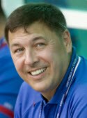
Rocco Monto, MD
(Photo by John Dorton, ISI)
Medscape: What brought you to this position?
Dr. Monto: I was a soccer player myself. I was a college All-American soccer player and played some professional ball before I went to medical school, so I've always had an interest in the game.
As a patient with many injuries during my career, going into orthopedics was natural. A lot of guys in my field are former athletes. It's what draws us to the field of orthopedics and sports medicine in particular.
Medscape: I was struck by how many names cropped up when I searched orthopedics, orthopedic physicians, and the Olympics.
Dr. Monto: You see a lot of them who are now productive orthopedic surgeons working in sports medicine. It's a natural fit for us as athletes. We know how to relate to athletic patients, and we know their sense of vulnerability. It really makes for a good match between doctor and patient.

Types of Injuries

Medscape: Could you describe the types of orthopedic injuries that are most common among soccer players?
Dr. Monto: Probably the most common injuries are the ankle sprain and the hamstring strain. There are very few players who can make it through a career without those injuries. After that would come fractures and typical lower extremity injuries. These are less common but can be more severe.
In soccer, we also have a lot of heading, and so concussive injuries and concussions have become a much more identified injury. It's probably no more common than it's always been, but we're identifying it with more prevalence now, and that's just because we're all tuned into the injury and the injury pattern more than we were before.
As for other injuries, surprisingly we see a lot of upper extremity injuries in soccer, usually from falls, whether it's shoulder dislocations or wrist injuries. After that are the ligament injuries, the anterior cruciate ligament (ACL) being the most common, particularly in female soccer players.
Medscape: Have you seen injuries that you would consider highly unusual in soccer?
Dr. Monto: I'm always surprised that we don't see more dental injuries than we do. I had my teeth knocked out as a college player. You would expect to see more than we actually do, with all the flying elbows and kicking that goes on. When we do see them, they can be quite severe. Most players don't wear mouth protection.

Avoiding Injuries

Medscape: When you're trying to teach people how to protect against these injuries, what kind of training is provided, or what do they have to do to make sure they're in condition to avoid an injury? I would imagine it's a matter of playing style but also certain types of strengthening.
Dr. Monto: The real revolution in soccer training in the past 15 years has been the addition of strength training. I think it's interesting that in our sport, there are many different philosophies, when you look at different countries, teams, and leagues and how they approach training. Despite those wide variations, however, the injury patterns remain fairly constant. Some of the things we can't change.
Things we have had success improving have been the incidence of ACL tears, particularly among women. Bert Mandelbaum, MD, of Santa Monica Orthopaedic and Sports Medicine Group, has done some fantastic work in helping women learn the risk factors that lead to ACL tears and the imbalance of the hamstring and quadricep muscles and how they land after they jump. Along with FIFA (Fédération Internationale de Football Association), our worldwide soccer group has developed training techniques to try to help the athletes avoid those injuries.
Medscape: Are there other Olympic competitions that have risks similar to soccer's?
Dr. Monto: It would be similar to men's team handball. I worked with the US handball team several years ago, and many of the injuries I see in soccer are similar to those in team handball. You see some of these in other sports as well, such as basketball.
A lot of the injuries that happen in soccer are noncontact. They happen away from the run of play, particularly ACL injuries. These injuries happen when you land after a jump or while trapping the ball, and there is a quick twist of the knee. The land-and-pivot problem is common to many sports.

Getting Back to the Field Sooner

Medscape: Are new therapies or techniques allowing soccer players to return to play earlier after an injury?
Dr. Monto: We've been much more open to the use of orthobiologic treatments, whether that's platelet-rich plasma treatments or more novel physical therapy approaches. We've gotten much better in getting our athletes back quicker, but as Freddie Fu, MD, in Pittsburgh, Pennsylvania, says, you can only heal so fast. We've pushed it without using any type of real medications or other potentially problematic techniques -- just using aggressive physical therapy and treatment and letting the body use its ability to heal. We're much better at doing that.
I'd say the biggest advance has been in using platelet-rich plasma and other types of treatments where we use growth factors to try to help people heal their muscle strains more quickly. In the past 2 years, that's been approved by the Olympic Committee and is now okay for use in Olympic athletes. It's not really a performance enhancer.
Platelet-rich plasma and bone marrow aspirate concentrate for more severe injuries are really the future in nonsurgical treatment of muscle strains, medial collateral ligament tears, and ankle sprains.

Bonding With the Athletes

Medscape: What about your experience as an athlete has enhanced your knowledge of orthopedics?
Dr. Monto: As an athlete who's become an orthopedic surgeon, I think the one thing that I took with me is the importance of the bond between the physician and the athlete and the importance of the personal relationship. A lot of trust is required, and gaining an athlete's trust is the most difficult part of being an orthopedic surgeon.
You really must have walked the walk to understand what athletes go through and the importance of decisions that might not be important to someone else. Whereas a doctor might feel that making next week's game isn't important, to an athlete it can mean the difference between completing a career successfully or not. You may be getting a player at the end of his career, and you need to understand how critical a little bit more time for him can be. You learn this eventually as a surgeon, but I think you learn it much earlier as an athlete.
Medscape: That presents an interesting dilemma.
Dr. Monto: Yes. This is where a surgeon has to have a very strong ethical and moral compass, because an athlete may be willing to take much higher risk than the surgeon will. This is where the bond and the trust come under a test, and you really have to put yourself in the athlete's shoes a little bit and understand where they're coming from, and also you have to communicate with them where you are too.
Obviously, no one wants to send an athlete to one last game that ends with them unable to walk right the rest of their lives. Nobody wants to put anyone in those kinds of precarious positions -- but again, it's all about relative value. Just as it's important to get a carpenter back to work as quickly as possible, it's also important to get the athlete back.
Then we have the pressures from fans, the media, owners, players, and other various interests -- sponsors and things like that, especially with the professionalism now that's pervasive in all the Olympic sports and test sports (women's boxing this year, and golf and rugby sevens to be added in 2016). These are all competing interests that you have to take into account to come up with the best compromise for the athlete.
Medscape: What do you most enjoy about being part of the US Soccer Team Physicians?
Dr. Monto: My favorites have always been the under-17 men's teams, because they are the stars of tomorrow. They still have an innocence and joy about the way they play and the way in which they approach the game and life, and it's always fantastic. It's just a real charge to work with those guys.
I remember being the doctor when Landon Donovan was a 16-year-old just making his way and suddenly we're playing in the Junior World Cup. [Landon Donovan is on the Los Angeles Galaxy team and is one of the world's most highly paid soccer players.] Those are fantastic experiences. They're very important to those players at that age, because they carry them for the rest of their career.
People don't quite understand how integrated the team physician is into the team and into the character and the fabric of the team. We're on the sidelines. We're with them in the games. We're with them in training. When you're taking care of the young athletes like they're children, they're part of your family, and that bond really helps them through crises when they get hurt. Those are things that people don't see. There's another whole layer of care for the athletes.

Olympics 2012: Nutrition Advice for the Athletic Patient

Olympics 2012: Nutrition Advice for the Athletic Patient

Olympics 2012: Nutrition Advice for the Athletic Patient

Marrecca Fiore; Nancy Clark, MS, RD, CSSD

Editor's Note:
With the 2012 Olympic games in full swing, it's a good time to review how best to advise the athletic patient on proper nutrition. Medscape interviewed Nancy Clark, a registered dietician and author of Nancy Clark's Sports Nutrition Guidebook, who offered advice on how to set up nutrition plans for athletes. A board-certified specialist in sports dietetics, Clark's clients have included players from the Boston Red Sox and Boston Celtics, as well as elite and Olympic athletes from a variety of sports.
Medscape: What are the first steps a clinician or healthcare provider should take in setting up a nutritional plan for an athletic patient?
Ms. Clark: The first thing I do is figure out their protein needs because protein needs are based on body weight. Athletes need about 1.2-1.7 g of protein per kilogram or 0.5-0.8 g protein per pound of body weight. From there, I figure out what the rest of their caloric needs are and fill in the plan with fruits, vegetables, and grains to make a balanced diet.
I advise even fueling throughout the day. Generally when I work with clients, I give them 4 food buckets. Every 4 hours they have a food bucket, so they are always fueling up or refueling. The food buckets are their breakfast, lunch one, lunch two, and dinner. If they train in the morning then they have part of their breakfast bucket before they work out and then the rest of it afterwards. If they are training in the afternoon, they might divide up the lunch one or lunch two buckets so that they are fueling and refueling around that training session. Regardless of when they work out, the plan evens out throughout the day so there is always a constant infusion of protein to build and repair muscles and carbs to fuel the muscles.
Medscape: Are the nutritional needs different according to the age and sex of the patient you're working with?
Ms. Clark: For certain, a 200-lb athlete has different needs from a 100-lb gymnast. But even though their food plates might look a lot different, they still have similar protein needs based on their body weight. Their calorie needs would vary. Their fluid needs would vary according to their body size. They just need different quantities of food.
Medscape: How should a provider determine how many calories a person needs? Is it just based on body weight? Or is it a combination of body weight and the type of athletic activity the patient is involved in?
Ms. Clark: I look at how many calories they need to breathe, which is their resting metabolic rate. Then I look at what they do when they're not training. Many athletes are very sedentary, so there is something called sedentary athlete syndrome. If they are doing double workouts, which a lot of them do, they train in the morning and then they lounge around and rest and recover. Then they train in the afternoon and then they lounge around and rest and recover. So even though they are training hard, when they're not training they're doing nothing and that can certainly affect their energy needs. That's the sedentary athlete as opposed to the athlete who trains and is not sedentary. That person trains and then is taking care of a family and doing the gardening, the laundry, the food shopping, and bringing the groceries in.
Medscape: How should providers figure out how much protein an athlete needs as opposed to their carbohydrate or healthy fat needs?
Ms. Clark: They need about 1.2-1.7 g of protein per kilogram or 0.5-0.8 g of protein per pound of body weight. Most people are already getting that, so it's a matter of distributing it evenly throughout the day, because generally a breakfast might be a bowl of oatmeal and dinner would be 3 chicken breasts. But I want them to more evenly divide their protein throughout the daytime so that they have protein with their oatmeal, or maybe instead of oatmeal they have some Greek yogurt with some nuts and toast with peanut butter, or they have a couple of poached eggs or some cottage cheese and fruit; this helps them even out how much protein they are eating. That is important for athletes who are weight conscious, because protein is very satiating, but also for athletes who want to optimize their muscle development and repair.
Medscape: Intense exercise tends to make people really hungry, so what kind of advice should physicians and healthcare providers give athletes so that they don't put on unwanted weight while training?
Ms. Clark: That is a common problem, particularly in women, because women tend to get hungrier than men do. This is where they really need to make sure they have protein at each meal and that they eat evenly throughout the day to prevent hunger. A lot of weight-conscious athletes will diet at breakfast and diet at lunch and then train on empty. Later, they end up starving and they blow it in the afternoon or evening because they have become too hungry; so, again we have to look at meal timing. Have them fuel by day and then lose weight at nighttime when they are sleeping. But they don't want to try to lose weight when they're training.
Medscape: And what advice should be given when the opposite happens and an athlete experiences unwanted weight loss while training?
Ms. Clark: I look at what they are drinking for fluids. Most of them are drinking a lot of water, and I just have them trade that water in for some kind of a healthy juice. Maybe they have more orange juice or grape juice or some low-fat chocolate milk. Juices and milk are 90%-95% water, but there is also some energy in it, and that energy adds value to fuel their muscles and be an additional source of calories to their sports diet.
Medscape: Why is eating so important to an athlete? How can it make or break somebody's performance?
Ms. Clark: Well, how important is gas to a car? You have a car; you put gas in it and it goes. You have a body; you put food in it and it goes a lot better. Certainly, it enhances stamina and endurance. Food is the sparkplugs that are needed for health, with the vitamins and minerals and the chemicals that fight inflammation.
Medscape: Olympic swimmer Ryan Lochte said in a recent interview that he ate poorly during the Beijing Olympics, adding that he ate at McDonald's almost every day. He mentioned this because he's since removed junk food from his diet with the hope of enhancing his performance. What type of effect does eating poorly have on an athlete's performance? Some of these Olympic athletes seem to be able to eat junk food but maintain a healthy body weight. Are these foods still damaging their bodies on the inside, even if you don't see any damage on the outside?
Ms. Clark: Definitely. If an athlete is filling up on Big Macs and French fries and fatty, greasy foods, the fat will fill the stomach but the muscles will remain unfueled. Only carbohydrates get stored in the muscles as glycogen; depleted muscle glycogen is associated with fatigue. You can go to McDonald's and you can get oatmeal, English muffins, juice, and fruit parfaits, so you can get a healthy carbohydrate-based diet if you look for it. But you can also go and choose the totally wrong things, and if your muscles aren't well fueled day after day after day, then you just get increasingly tired. When you are trying to perform at your best, you really want a foundation of healthy carbs at each meal.
But just as food can be powerfully bad for you, it can be powerfully good for you. If you put quality, premium nutrition in your tank, it makes a big difference -- not just in terms of energy level, but also in terms of health and vitality.
Medscape: How should clinicians advise patients who aren't currently athletic but are inspired by the Olympics or some other event to begin working out? How should they approach their nutritional needs?
Ms. Clark: I start at breakfast. As I mentioned, if you have a car, you put gas in it and it goes, so you want to have a quality breakfast so you can go. Research suggests that people who have a high-protein breakfast end up eating fewer calories at the end of the day. If you have a dinner-size portion of protein at breakfast, it feeds you throughout the day and it keeps you fed so that it is easier to bypass the doughnuts, the Danish pastry, and the so-called junk food that manages to creep into people's lives when they haven't had much of a good breakfast. If you start out with a substantial breakfast, you'll have good energy, and it keeps you satiated so you feel like going to the gym. Even at the end of the workday you'll still have some energy to go to the gym.
With the workout, the place to start is to do some strengthening exercises to strengthen your muscles. If your muscles are stronger, it's easier to walk farther, run farther, bike farther. But first you get stronger and then you add the more aerobic exercise. Unfortunately most people start out with, "Oh, I'm going to run a mile," but it's much more important to get strong first.
The Olympics is a great time for people to take a look at how sedentary they are, how they could get in shape even if they just got up and marched in place during TV commercials, or if they kept little weights by their chair and got up to lift some weights a couple of times each day. There is a lot that can be done if people get creative and just figure out how they can move their bodies more. And certainly sitting around, even sitting all day at work, is an occupational health hazard. We know that smoking and being around smoke is a health hazard, but if people look at sitting as being hazardous to their health as well, then they can take small steps to move a little more.